Feline Infectious Peritonitis (FIP) is a fatal disease caused by certain mutant strains of feline coronavirus (FCoV).The pathology of FIP can be characterized according to its two main forms- -wet (exudative type) and dry (non-exudative type)
Wet FIP (exudative type)
Wet FIP is characterized by the accumulation of fluid in body cavities, usually in the abdominal and thoracic cavities. These effusions are usually:
Yellow to clear fluid that may be viscous.
High protein content (total protein concentration usually greater than 3.5 g/dL).
Low cellular content, with neutrophils and macrophages predominating.
Pathologic features
Vasculitis: wet FIP often presents with perivascular inflammation called necrotizing vasculitis. The walls of the blood vessels are eroded, leading to accumulation of exudate.
Plasma membrane inflammation: the peritoneum, pleura, and pericardium often show inflammation in the form of fibrin deposition and inflammatory cell infiltration.
Dry FIP (non-exudative type)
Dry FIP is characterized by granulomatous lesions of multiple tissues and organs rather than body cavity fluid collection. These granulomas are found primarily in: Liver, kidneys, lymph nodes, spleen, CNS
Pathologic features
Granulomas and nodules: granulomatous lesions formed by macrophages, lymphocytes, and plasma cells, commonly found in the above organs.
Inflammatory response: infiltration of inflammatory cells, predominantly monocytes and macrophages, accompanied by tissue necrosis and fibrosis.
CNS lesions: inflammatory cell infiltration can be seen in the meninges and brain parenchyma, manifesting as meningoencephalitis.
Common pathologic features
Both wet FIP and dry FIP share the following common pathologic features:
Systemic inflammatory response: extensive immune-mediated inflammatory response due to intravascular deposition of immune complexes.
Multisystemic involvement: extensive involvement of different organ systems, including the digestive system, respiratory system, and nervous system.
Lymphadenopathy: lymph nodes are often enlarged and show nonspecific inflammation and hyperplasia.
Diagnostic basis
The definitive diagnosis of FIP relies on pathologic tests, including:
Tissue biopsy: Pathologic examination of tissue sections is the gold standard for confirming the diagnosis of FIP. Typical pathologic features include vasculitis and granulomatous lesions as described above.
Immunohistochemical staining: can be used to detect FCoV antigen and help confirm the diagnosis.
Because of the diverse clinical presentation of FIP, confirming the diagnosis usually requires a combination of clinical signs, laboratory tests (e.g., ascites analysis, serum protein electrophoresis, etc.), imaging studies, and pathologic findings.
Feline Infectious Peritonitis (FIP) is a fatal disease caused by feline coronavirus (FCoV).FIP can be categorized as wet or dry, and the symptoms are complex and varied, making diagnosis difficult. The main points involved in the question of whether to perform antigen testing are as follows:
The role of antigen testing
Antigenic tests (e.g. ELISA, IFA, etc.) are primarily used to detect the presence of FCoV in cats.It is important to note, however, There are two forms of FCoV: a relatively harmless enteric FCoV (FECV) and an FIP-causing FCoV (FIPV). Antigen testing cannot distinguish between these two forms, so a positive test for FCoV does not necessarily mean that a cat has FIP.
Since most cats will contract and carry FCoV throughout their lives (especially cats living in multi-cat environments), a diagnosis of FIP based on a positive antigen test result alone is not accurate.
Testing for fip in cats
CLINICAL symptoms: Diagnosis of FIP is usually based on clinical symptoms, history, and physical examination. Wet FIP results in an accumulation of fluid in the abdominal or thoracic cavity, while dry FIP presents as nodules in internal organs.
Laboratory tests: Ancillary tests such as complete blood count, serum protein electrophoresis, and analysis of ascites or pleural fluid can provide further information.
PCR assay: Polymerase chain reaction (PCR) assay can be used to detect RNA of FCoV, which helps to confirm the diagnosis of FIP, but its sensitivity and specificity need to be further verified.
Tissue biopsy: In some cases, the diagnosis of FIP can be confirmed by tissue biopsy and pathology.
Antibody test vs Antigen test for fip
Antibody test: used to detect the presence of antibodies against FCoV in cats. This test also cannot distinguish between FECV and FIPV, but can provide information on whether the cat has been exposed to FCoV.
Antigen Test: Used to directly detect the presence of the virus and, as mentioned earlier, has limited diagnostic significance due to the inability to differentiate between the different forms of FCoV.
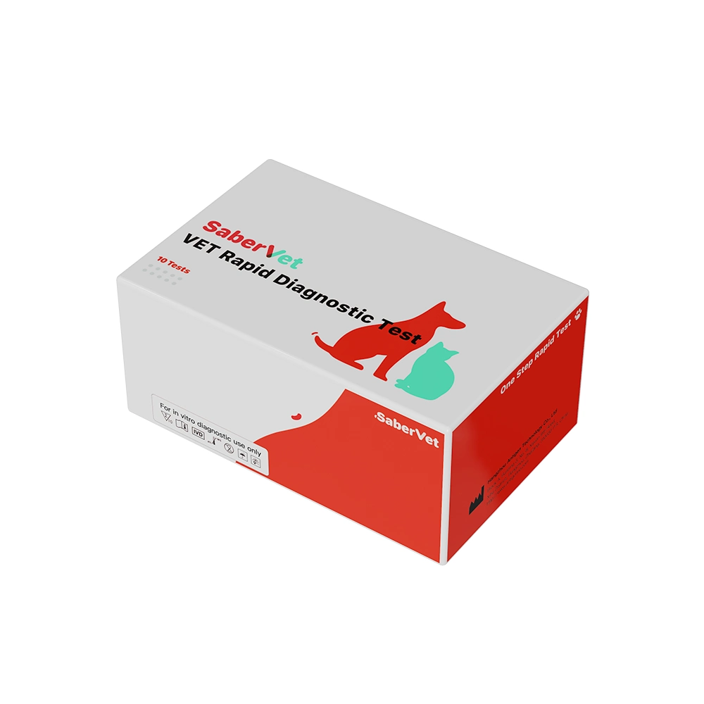
The SaberVet Pet Rapid Test Kit, the pioneer in animal diagnostics. Our innovative technology and rapid diagnostic products create accurate and timely health protection for your pet’s health.


